News
Notice of Publication of the Results on Detection of Breast Cancer in Histopathological Micrographs Using Single-Shot Multi-box Detector (SSD)
Renascience endeavors to solve issues of medical or social importance, and especially focuses on medical issues related to aging population combined with the declining number of children, as well as research and development on women’s and children’s diseases.
As one sphere of research and development in the area of women’s diseases, a paper on detection of breast cancer in histopathological micrographs using a single-shot multi-box detector (SSD)*1, an object detection method using artificial intelligence (AI), has been published in the peer-reviewed scientific journal “Journal of Pathology Informatics” as the result of the collaboration with Professor Takashi Suzuki of the Department of Pathology and Laboratory Medicine, Tohoku University Graduate School of Medicine.
Yamaguchi M, Sasaki T, Uemura K, Tajima Y, Kato S, Takagi K, Yamazaki Y, Saito-Koyama R, Inoue C, Kawaguchi K, Soma T, Miyata T, Suzuki T
Automatic breast carcinoma detection in histopathological micrographs based on Single Shot Multibox Detector Journal of Pathology Informatics Volume 13, 2022, 100147
(https://www.sciencedirect.com/science/article/pii/S2153353922007416?via%3Dihub)
Diagnosis by histological classification by pathologists is critical for appropriate treatment to improve the prognosis of breast cancer patients. However, since the number of pathologists is limited, pathological diagnosis supported by artificial intelligence (AI) or other means is important. Therefore, we have developed a model for automatic detection of breast lesions in microscopic histopathology images using a single-shot multi-box detector (SSD) and evaluated the image diagnosis support function by AI.
After building a dataset and training the SSD model on 1,361 microscopic images, we used 315 images to evaluate the difference in pathology diagnosis between pathologists and medical students with and without SSD model use.
The model for the diagnosis support achieved diagnostic accuracy of 88.3% and 90.5% when attempting to classify 3 classes (benign, non-invasive*2, or invasive cancer*3) or 2 classes (benign or malignant), respectively, and IoU (Intersection over Union) *4, the evaluation index in object detection, was 0.59. The classification performance by the medical students was extremely high, with an average diagnostic accuracy of 84.7% using the model, compared to 67.4% without the model.
*1: Single-shot multi-box detector (SSD)
A type of object detection model. Unlike the object detection model with conventional convolutional neural network (CNN), each CNN operation simultaneously “detects the regions of interest” and “classifies the object”. This makes it possible to speed up the process for the object detection.
*2: Non-invasive cancer
In breast cancer, the non-invasive cancer means the cancer still in the ducts and lobules where it first develops.
*3: Invasive cancer
In breast cancer, invasive cancer means the cancer that has spread beyond the ducts and lobules into the stroma outside the ducts.
*4: IoU (Intersection over Union)
It is an index to evaluate the amount of error between the bounding box of the object area predicted by the object detection model and the correct bounding box. It is simply called “Intersection over Union”, since it is the ration obtained by dividing Intersection with (over) Union to evaluate the degree of the overlap between the boxes.
Example images of detection by pathologists
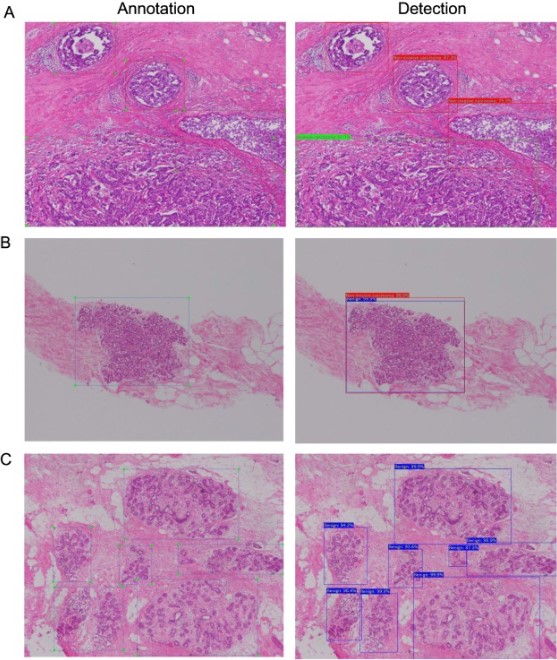
Example of annotation (left) and detection by the model (right). Blue, red, and green boxes indicate benign, noninvasive, and invasive cancers, respectively.
a: The model accurately detected the cancers.
b: images that contain boxes detecting different labels in the same region.
c: The model accurately detected benign regions with bounding boxed of different shapes from the annotation.