News
Publication of a paper on artificial intelligence for breast cancer detection in histopathological images during intraoperative rapid diagnosis
Our company is engaged in research and development to address important medical and social issues, such as the declining birthrate and aging population, including diseases affecting women and children. In collaboration with Professor Takashi Suzuki and Assistant Professor Mio Tanaka of the Department of Pathology, Graduate School of Medicine, Tohoku University, and NEC Solution Innovators, Ltd. (NES), we have been developing artificial intelligence (AI) to support intraoperative rapid diagnosis2) of breast cancer using frozen pathological sections1). We are pleased to announce that the results of this collaborative research have been published in The Tohoku Journal of Experimental Medicine as a paper titled “Automatic Breast Carcinoma Detection in Histopathological Micrographs Based on Single Shot Multibox Detector3).”
Yamaguchi M, Sasaki T, Uemura K, Tajima Y, Kato S, Asakura T, Takagi K, Yamazaki Y,Masamune A, Miyata T, Suzuki T.Artificial Intelligence for Breast Carcinoma Detection in Histopathological Images Based on Single Shot Multibox Detector in Intraoperative Rapid Diagnosis, J-STAGE, Article ID: 2025.J120
( https://www.jstage.jst.go.jp/article/tjem/advpub/0/advpub_2025.J120/_article/-char/en )
Background
Breast cancer is the most prevalent cancer among Japanese women, with one in 11 women estimated to suffer from breast cancer in their lifetime. When breast cancer is suspected based on a lump or diagnostic imaging, the final diagnosis is a pathological diagnosis. Accurate diagnosis based on histological classification by a pathologist is essential for prompt and appropriate treatment of breast cancer. However, due to the limited number of pathologists, there is a growing need for AI-based pathological diagnosis support.
In collaboration with Tohoku University and others, we are developing AI to detect breast cancer lesions from pathology images. On October 7, 2022, we announced that the results of our joint research on a breast cancer detection model in histopathological micrographs4) using a single-shot multibox detector (SSD), an AI-based object detection method, were published in the the Journal of Pathology Informatics. When the detection model classified cancers into three classes (benign, non-invasive cancer, invasive cancer) or two classes (benign, malignant), the accuracy was 88.3 % and 90.5 %, respectively.
In the current study, we applied this technology to intraoperative rapid pathological diagnosis of breast cancer. Rapid intraoperative diagnosis is extremely important for determining the extent of breast cancer surgery, but it requires pathologists with high expertise in breast cancer pathological diagnosis. A diagnosis must be made within 10–20 minutes of receiving the specimen, and the quality of frozen sections is often poor. To address these challenges, we have been developing AI to support rapid intraoperative diagnosis.
Results
After learning data consisting of 943 microscopic images of intraoperative frozen sections of breast cancer3), an SSD model was developed. The diagnostic accuracy of breast cancer was evaluated using 65 intraoperative frozen section images.
- Breast cancer classification: benign, malignant (non-invasive cancer4), and invasive cancer5))
- Diagnostic accuracy:
Benign/malignant classification: 92.3 % accuracy rate
Benign/malignant classification plus cancer classification (non-invasive cancer/invasive cancer): 89.2 % accuracy rate - Average detection time: 0.875 seconds per image
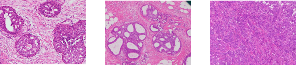
benign non-invasive cancer invasive cancer
This AI-based diagnostic technology enables support for rapid intraoperative diagnosis of breast cancer, suggesting its potential as an effective tool for reducing the burden on pathologists and contributing to surgical support through intraoperative diagnosis.
1) Frozen Pathological Section
A frozen pathological section is a method in which tissue removed from a tumor, such as breast cancer, during surgery is rapidly frozen, thinly sliced, and examined under a microscope for diagnostic purposes. This technique allows surgeons to quickly determine whether the tumor has been completely removed and whether cancer has spread to the lymph nodes, enabling them to make immediate decisions regarding additional tissue removal or other surgical actions if necessary.
2) Intraoperative rapid Diagnosis
A method for diagnosing cancer by freezing tissue removed during surgery and quickly observing it under a microscope.
3) Single-Shot Multibox Detector (SSD)
A type of image detection AI. In this study, it detects and classifies breast cancer lesions from microscopic images of intraoperative frozen sections.
4) Histopathological micrographs
A histopathological microscopic image is created by thinly slicing and staining tissue, such as breast cancer tissue or lymph nodes, removed during surgery and then capturing it under a microscope. Observing the tissue in this way allows for the assessment of the presence of cancer cells, their morphology, growth patterns, and the extent of spread to surrounding tissues. This provides important information that helps evaluate the effectiveness of the surgery and guides decisions regarding further treatment.
5) Non-invasive Cancer
Cancer in which cancer cells have not penetrated the basement membrane into the surrounding stroma. While the risk of metastasis to surrounding tissue is low, confirmation of the tumor border is important during intraoperative diagnosis.
6) Invasive Cancer
Cancer in which cancer cells have penetrated the basement membrane into the surrounding stroma. There is a possibility of vascular or lymphatic invasion and metastasis, making this important for determining the surgical procedure and extent of resection.